
Cadaver lab provides med students with ‘first patients,’ invaluable training


Weighing just 90 pounds, the 90-year-old female on the table has a long medical history, including multiple broken bones, cataract removal and a hysterectomy, as well as the metastasizing kidney cancer and COPD that killed her.
The body lies face up, covered from head to toe in a white sheet, a medical history tacked to the laboratory wall.
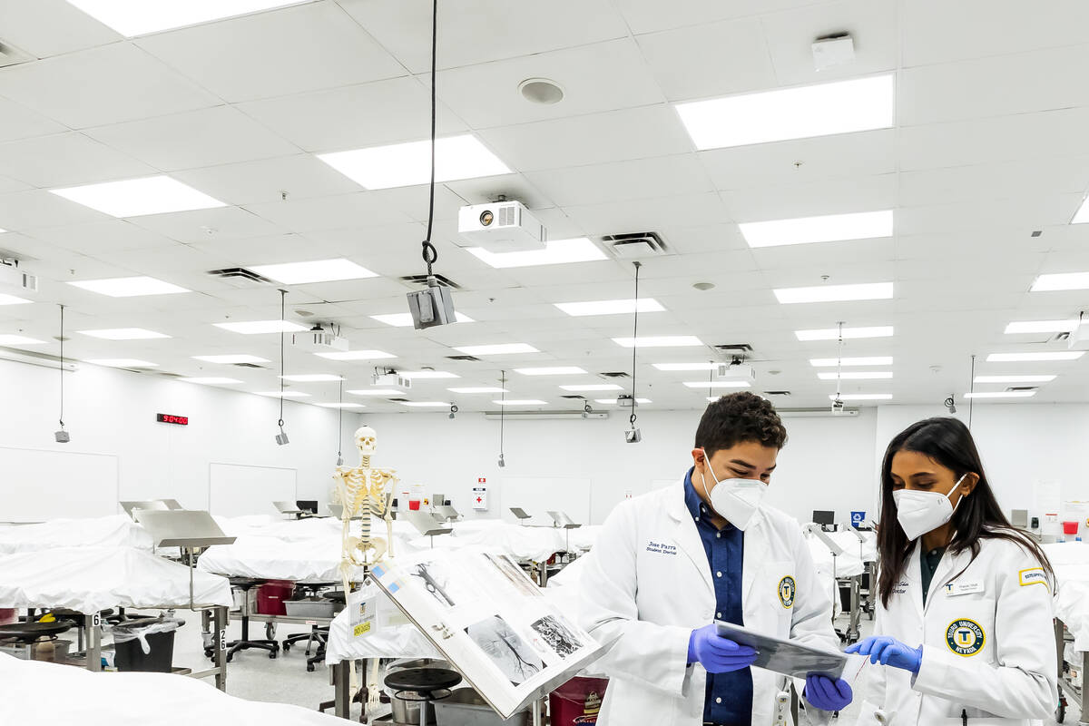
It is one of 40 bodies in Touro University Nevada’s cadaver lab, the only facility of its kind in Southern Nevada. There, first-year medical students learn about anatomy from the cadavers they consider their first patients.
At a time when anatomy increasingly is taught using interactive touch screens and anatomical models instead of scalpels and cadavers, the university’s leaders continue to believe in the importance of learning by dissecting actual bodies, medicine’s centuries-old rite of passage.
Despite technological innovations, “the so-called psychomotor skill — the ability to use your hand to dissect, to cut and feel — was found to be really, really important in a student’s ability to learn about the body,” said Dr. Wolfgang Gilliar, dean of the College of Osteopathic Medicine at the university in Henderson. “You really learn the 3D relationships by having a real cadaver.”
Second-year student Pranati Shah, who wielded a scalpel for the first time in the lab, said cutting into actual tissue has given her a better grasp of anatomy.
“A nerve and an artery and a vein sometimes look exactly the same,” the 24-year-old said. “And so I think the hands-on here makes a huge difference. When you press up on a vein, you’re like, ‘This is collapsing. This is a vein.’ When you press on a nerve, it’s like, ‘This is harder. This is a nerve. I don’t want to cut this.’”
She said it’s been instructive to see abnormalities in cadavers that are a far cry from the perfect anatomical structures laid out in textbooks. Having found a hernia in her cadaver, Shah said, “Now I’m never going to forget what a hernia looks like in my entire life because I kind of discovered that in my body on my own.”
Third-year student Jose Parra, 26, said, “I have my cadaver in my brain. … When you go in and do these dissections, when you see something like that, you don’t forget.”
Touro medical students study anatomy in the lab during the second semester of their first year of medical school.
Four to six students are assigned as a team to a single cadaver for the semester. Students also may study the cadavers assigned to other teams, as no two bodies are identical, the students said.
Each cadaver lies face up on an individual table. Beneath the table are buckets, saws and other tools needed for dissection. Overhead are video cameras. On the walls are large screens.
The temperature in the lab is a slightly chilly 65 degrees, and even with an advanced air filtration system, there is a slight scent of the formaldehyde used to preserve the bodies.
Despite the somber environment, Shah said the lab became the focal point of both her academic and social life, a place where friendships blossomed. Touro continued using the lab during the pandemic by spacing tables and students farther apart.
“It’s easy to feel isolated during medical school by itself, and then with COVID, too, it’s like, how am I going to make friends and kind of create a support system?” she said. “It’s crazy to think that this room did that, but it really did.”
Virtual anatomy labs
Yet some medical schools are moving away from traditional cadaver labs, saying they take up too much of a student’s time and a school’s resources.
But UNLV’s Kirk Kerkorian School of Medicine, which opened in 2017, met with considerable pushback over its initial plan to use only advanced technologies to teach anatomy in place of a cadaver lab, said Dr. Neil Haycocks, vice dean for academic affairs and education.
It revised its plans to include an eight-table lab, which Haycocks expects to be completed this year, where faculty can demonstrate dissection to students. The school also offers an elective dissection course for fourth-year students in a rented medical training space.
“I think we’re getting the best of both worlds,” he said.
The amount of knowledge that medical students need to learn is increasing exponentially, he said. As a result, UNLV is putting a greater emphasis now on “clinically relevant problem-solving,” Haycocks said.
“We think by removing the dissection component, that just basically gives the students a much more efficient and effective learning experience overall,” he said.
Roseman University of Health Sciences in Southern Nevada, which is fundraising for a medical school, has pivoted from plans for a cadaver lab, said Dr. Karin Esposito, the college of medicine’s senior executive dean for academic and student affairs. It now favors using plasticized, pre-dissected specimens and augmented and virtual reality teaching tools, which unlike a cadaver, a student can return to again and again for training, she said.
“The augmented and virtual reality tools are getting to the point where they are probably going to be surgical training tools very soon,” she said. “In some cases, they probably already are. So surgeons can take 3D images that are potentially even reconstructed from the actual patient that they’re going to operate on — from MRIs, etcetera — and walk themselves through the surgery. That’s how precise that’s getting.”
Shelley Berkley, CEO and senior provost at Touro University Nevada, said advanced technologies can complement cadaver dissection but can’t take its place. The university gave a conservative estimate of $250,000 as the annual cost of operating the lab.
“As long as I’m at Touro, we will continue to have cadavers,” said Berkley, an attorney and former Nevada congresswoman.
“As technology becomes more sophisticated and more available, we’ll of course incorporate that technology into our training. But in the end, there’s nothing like hands-on getting into the body and seeing how it really looks,” she said. “And at this moment in time, as sophisticated as the technology has become,I still don’t think it’s a substitute for the body.”
White rose ceremony
Touro obtains its cadavers from the University of North Texas. Bodies used as cadavers may have been donated by individuals for scientific purposes or may have been unclaimed. Touro does not itself accept body donations.
Touro, a Jewish university, will not provide to its students for dissection bodies with offensive tattoos, such as those with anti-Jewish slurs or symbols, said Jeremy Day, the lab’s manager.
The university instills in its students a respect for the bodies, emphasizing that they are the students’ first patients, both students and faculty said.
At the end of the school year, Touro faculty and students hold a white rose donor appreciation ceremony described as a solemn and sometimes emotional affair. The bodies are then cremated and the ashes returned to the University of North Texas, which in turn provides the remains to donors’ families.
The students are “very appreciative of that first patient and the knowledge they acquire,” Berkley said. “And consequently, when we do the white rose ceremony at the end of the year, our students are essentially thanking their first patients for the knowledge that they’ve acquired.”
Contact Mary Hynes at mhynes@reviewjournal.com or 702-383-0336. Follow @MaryHynes1 on Twitter.


